
Meibomian gland dysfunction (MGD) is a chronic problem with the oil glands in the eyelids, usually from thickened oil and blocked gland openings. It is one of the most common causes of evaporative dry eye. At Copper View in South Jordan, MGD is evaluated with gland expression and meibography and treated with home care, prescriptions, and in-office options such as IPL, radiofrequency, low-level light therapy, and gland probing.
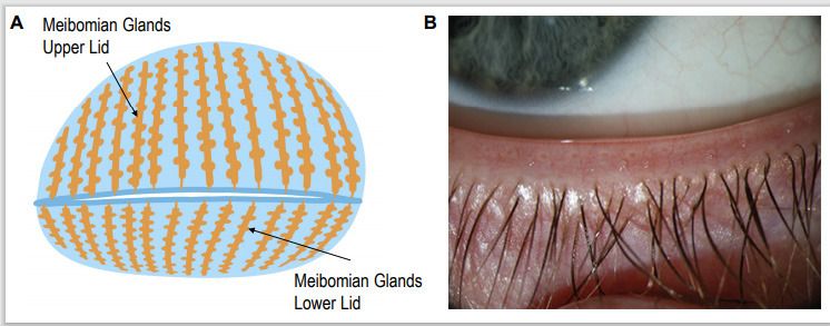

How meibomian glands work
Each upper eyelid has roughly 25 to 40 meibomian glands, and each lower lid roughly 20 to 30. They run vertically inside the lid, like the teeth of a comb, and open in a single row just behind the lashes.
Every blink squeezes a small amount of oil, called meibum, onto the lid edge. The blink then spreads it across the tear film as a very thin top layer. That oil layer slows evaporation and keeps the tears smooth enough to see through clearly.
Healthy meibum is clear and flows like light olive oil at body temperature. In MGD, it becomes cloudy, then thick like toothpaste, and eventually may not come out at all.
What goes wrong in MGD
The most common form is obstructive MGD. The cells lining the gland opening overgrow and plug the duct, and the oil itself gets thicker. Backed-up glands become inflamed, and over time the gland tissue can shrink. This is called gland dropout or atrophy.
When the oil layer thins, tears evaporate quickly. The remaining tears become saltier, which irritates the surface and triggers inflammation. That inflammation then makes the glands worse. This loop is why MGD is the leading cause of evaporative dry eye.
Things that make MGD more likely
- Age, and hormonal changes including menopause
- Rosacea, including ocular rosacea
- Demodex mites on the lashes
- Long screen sessions with reduced or partial blinking
- Contact lens wear
- Some medications, such as isotretinoin for acne
- Eyeliner and makeup on the lid margin
- Low-humidity environments, which describes much of Utah
Symptoms
MGD symptoms often look like “dry eye,” but some patterns point toward the glands:
- Burning or grittiness that gets worse through the day
- Vision that blurs and sharpens with blinks
- Watery eyes, especially outdoors or in wind
- Red, thickened, or crusty lid edges
- Recurring styes or chalazia
- Contacts that feel fine in the morning and terrible by afternoon
How we diagnose MGD
Depending on your symptoms and exam findings, an MGD evaluation may include:
- Lid margin exam at the slit lamp, looking for capped gland openings, irregular lid edges, and abnormal blood vessels
- Gland expression, gentle pressure to see what the glands release and how many respond
- Tear breakup time, which is usually fast when the oil layer is poor
- Meibography, infrared imaging that shows gland length and dropout
- Inflammation testing such as MMP-9 when the result would change the plan
- Screening for Demodex and signs of ocular rosacea
Seeing your own meibography images is often the moment MGD makes sense. You can see whether glands are full length, shortened, or missing, and we can compare images over time.
Why haven’t warm compresses fixed it?
This is one of the most common questions we hear. Warm compresses are reasonable advice, but they have limits:
- They cool off fast. Meibum in MGD may need sustained heat to soften. A washcloth loses most of its heat in a minute or two.
- They can’t open scarred openings. If the duct opening is sealed by tissue, heat alone won’t clear it.
- They don’t treat inflammation. Abnormal blood vessels along the lid, ocular rosacea, and Demodex keep driving the problem.
- They don’t help tear production. If you also have aqueous-deficient dry eye, the oil layer is only part of the story.
A good heated mask, used consistently, is still a helpful foundation. For moderate or advanced MGD, it’s usually one layer of a broader plan.
Treatment options for MGD
Treatment is chosen based on how blocked the glands are, how much inflammation is present, and what else is going on.
| Treatment | What it targets | Typical role |
|---|---|---|
| Lid hygiene and heat | Debris, bacteria, mild oil thickening | Foundation for almost everyone |
| Prescription drops and oral medications | Inflammation, Demodex, evaporation, rosacea-related changes | Common early step |
| IPL | Abnormal lid blood vessels and inflammation, gland function | MGD with inflammation or rosacea |
| Radiofrequency | Deep, sustained eyelid heating to soften oil | Thickened meibum, often with expression |
| Low-level light therapy | Gentle lid warming and light exposure | Alone or combined with other treatments |
| Gland probing | Physically opens obstructed or scarred gland ducts | Glands that do not respond to heat and expression |
In-office treatments are usually given as a series, often with gland expression at the same visit. We recheck symptoms, tear breakup time, and gland function to decide whether more sessions or maintenance visits make sense.
Can damaged meibomian glands grow back?
Here is the careful answer. Gland tissue that has completely atrophied is generally considered lost, and no current treatment reliably regrows it. Glands that are shortened, plugged, or underperforming can often function better once obstruction and inflammation are addressed.
A few published case series after gland probing have described apparent increases in gland tissue on meibography. Those findings are interesting but come from small studies, and regrowth is not a result anyone should promise.
What that means for you: the glands you have now are worth protecting. Treating MGD earlier, before more dropout occurs, generally gives the best chance of keeping eyes comfortable.
Living with MGD
MGD is chronic for most people. Good management usually combines:
- A home routine you will actually keep, including lid hygiene and heat
- Blink breaks during screen work
- A humidifier at home in winter, which helps in the Salt Lake Valley
- Careful choices about eyeliner, makeup, and skincare near the lids
- Periodic in-office maintenance if it has helped you
Dr. Simmons and our team will help you figure out which parts make the biggest difference for your eyes.
Frequently asked questions
Can damaged meibomian glands grow back?
Glands that have fully atrophied are generally considered permanent loss, and no treatment reliably regrows them. Glands that are shortened, blocked, or underperforming can often work better once obstruction and inflammation are treated. Some small studies after gland probing have reported apparent regrowth of gland tissue on meibography, but this is not established as a predictable outcome. The practical message: protect and treat the glands you have, and earlier is better.
How do I know whether my meibomian glands are blocked?
You can't tell for sure at home. Clues include burning, grittiness, blurry vision that clears with blinking, eyes that water in wind, crusty or red lid edges, and styes or chalazia. In the office, pressing gently on the lid shows whether the glands release clear oil or something thick, and meibography images show the glands' structure.
Why haven't warm compresses fixed my dry eye?
Most warm compresses cool off within a minute or two, which is not long enough to melt thickened gland oil. Compresses also cannot open scarred gland openings, treat lid inflammation, or address Demodex mites, rosacea, or tear production problems. They are a good foundation but are often not enough on their own for moderate or advanced MGD.
Is MGD the same as blepharitis?
They overlap but are not the same. Blepharitis means inflammation of the eyelid margin and can involve the lashes, bacteria, or Demodex mites. MGD is a problem with the oil glands themselves. Posterior blepharitis is often caused by MGD, and many patients have both.
Does MGD cause styes and chalazia?
A chalazion is a blocked, inflamed meibomian gland that forms a firm bump, and people with MGD tend to get them more often. Treating the underlying gland dysfunction may reduce how often they recur.
Is MGD treatment covered by insurance?
The evaluation and many prescriptions are often billed to medical insurance. In-office treatments such as IPL, radiofrequency, and low-level light therapy are commonly not covered. We review costs with you before treatment.
Sources and further reading
- Nichols KK, et al. The International Workshop on Meibomian Gland Dysfunction: Executive Summary. Investigative Ophthalmology & Visual Science. 2011;52(4):1922-1929.
- Nelson JD, et al. The International Workshop on Meibomian Gland Dysfunction: Report of the Definition and Classification Subcommittee. IOVS. 2011;52(4):1930-1937.
- Bron AJ, et al. TFOS DEWS II Pathophysiology Report. The Ocular Surface. 2017;15(3):438-510.
- Jones L, et al. TFOS DEWS II Management and Therapy Report. The Ocular Surface. 2017;15(3):575-628.
- Xue AL, et al. Randomised double-masked placebo-controlled trial of the cumulative treatment efficacy profile of intense pulsed light therapy for meibomian gland dysfunction. The Ocular Surface. 2020;18(2):286-297.
- Maskin SL. Intraductal meibomian gland probing relieves symptoms of obstructive meibomian gland dysfunction. Cornea. 2010;29(10):1145-1152.
- Maskin SL, Testa WR. Growth of meibomian gland tissue after intraductal meibomian gland probing in patients with obstructive meibomian gland dysfunction. British Journal of Ophthalmology. 2018;102(1):59-68.
- American Academy of Ophthalmology. What Is Blepharitis?